This web page was produced as an assignment for Genetics 564, an undergraduate course at UW-Madison
What are Model Organisms?
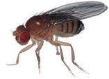
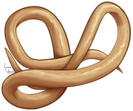
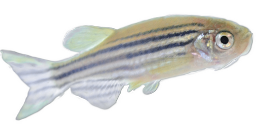
Model organisms are used to model and study human disease. Many studies cannot be done in humans for ethical reasons making model organisms essential to research. There are many factors to consider when choosing a model organism including genotypic and phenotypic similarity to humans, gestation time, cost, and east of working with. Common model organisms include mice, rats, fruit flies, roundworms, zebrafish and yeast, each with their own advantages and disadvantages.

|
|
|
|

|
|
Model Organisms and COL3A1
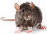
Of the aforementioned model organisms, COL3A1 homologs are present in mice and rats. While mice and rats are two of the more complex and expensive model organisms they are also very similar to humans which is evident in the 90% identity to humans obtained for both when using BLAST to compare homologs.
To find mutant phenotypes in mice and rats Mouse Genomic Informatics (MGI) and RGD databases were used respectively.
Mouse Models
|
COL3A1 mutations that were used in mouse models were chemically induced, spontaneous, or targeted. Targeted mutations were generated by creating knockout mice via homologous recombination. See video at right for more information on the generation of knockout organism. Along with removing gene function, a model or a transgene composed of COL3A1 regulatory segments fused to the reporter gene GFP exists. This model is useful to visually study the regulation of COL3A1 without disrupting function. |
|
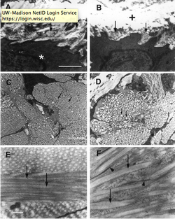
Figure 1: Phenotypes of COL3A1 knockout mice compared to wild type (WT)
A) Collagen fibrils of WT mouse aorta
B) Collagen fibrils are missing from aorta of COL3A1 mutant
C) Cross section of fibrils in WT adventitia, arrows pointing to individual fibrils
D) Diameter of fibrils in mutant adventitia is decreased
E) Collagen fibrils are uniform in WT skin sample
F) Collagen fibrils in skin are thicker in diameter and not uniform in mutant
Based on experimental data, COL3A1 mouse mutants typically die within 48 hours of birth. Phenotypically they have reduced body size, skin lesions, enlarged intestines, and irregular collagen structure (shown above). Those that do not die within 48 hours die by 6 months of age due to ruptured blood vessels or intestinal rupture (2).
Analysis
COL3A1 knockout mice exhibit phenotypes similar to human affected with Vascular EDS making mice a good model organism to study. Several experiments that could be done in mice to further our understanding of Vascular EDS include: knockdown genes that are found to play a role in hollow organ wall development from transcriptomic analysis, study the localization of COL3A1 using transgenic reporter genes, and determining the effect of overexpression of COL3A1.
References:
1) Liu X, Proc Natl Acad Sci U S A 1997 Mar 4;94(5):1852-6 SS 18 /C?. Copyright 1997 National Academy of Sciences, U.S.A. \
2)http://www.informatics.jax.org/image/MGI:3720373
1) Liu X, Proc Natl Acad Sci U S A 1997 Mar 4;94(5):1852-6 SS 18 /C?. Copyright 1997 National Academy of Sciences, U.S.A. \
2)http://www.informatics.jax.org/image/MGI:3720373
